Upper limb motor NCS
|
Nerve |
Origin |
Active |
Reference |
Ground |
Stimulation |
Lat. (Max) |
Amp. (Min) |
NCV (Min) |
F Lat. (Max) |
|
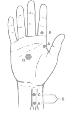
Median to Abd Pol
|
C8,T1 |
halfway between the midpoint of the distal wrist crease and the first MCP joint |
slightly distal to the first MCP joint |
Between active electrode and the cathode. |
A- The cathode is placed 8 cm proximal to the active electrode, slightly medial to the tendon of the flexor carpi radialis. B- medial to the tendon of the biceps at the elbow, slightly proximal to the antecubital crease |
4.5 |
4 |
50 |
32 |
|
Median
to Flex Carp Rad
|
C6,7,8 |
Over the belly of the flexor carpi radialis, one third of the distance from the medial epicondyle to the radial styloid. |
over the radial styloid |
on the dorsum of the hand. |
medial to the tendon of the biceps at the elbow, slightly proximal to the antecubital crease |
3.6 |
2.3 |
|
|
|
Median
to Flex Pol Long (ant interosseous)
|
C7,8,T1 |
On the lateral forearm, 10.2 cm from the distal wrist crease |
over the distal tendon of the flexor pollicis longus |
Between the stimulating and recording electrodes. |
medial to the tendon of the biceps at the elbow, slightly proximal to the antecubital crease |
4 |
2.5 |
|
|
|
Median
to Pron Quadrat (ant interosseous)
|
C7,8,T1 |
Midpoint between the radius and ulna on the dorsal forearm, 3 cm proximal to ulnar styloid |
over the radial styloid |
on the dorsum of the hand. |
medial to the tendon of the biceps at the elbow, slightly proximal to the antecubital crease |
5 |
1.5 |
|
|
|
Median
to Pron Teres
|
C6,7 |
An equilateral triangle is imagined, with the medial epicondyle and the biceps tendon (at the level of the epicondyle) as two of its points. The active electrode is placed at the third point, on the proximal forearm |
over the radial styloid |
on the dorsum of the hand. |
medial to the tendon of the biceps at the elbow, slightly proximal to the antecubital crease, 10 cm above active electrode. |
3.5 |
3 |
|
|
|
Median
to 1st Lumbrical
|
C8,T1 |
Placement is on the palm, slightly radial to the long flexor tendon of the index finger (localized by flexion of the index finger) and 1 cm proximal to the midpalmar crease |
at the base of the index finger |
Between active electrode and the cathode. |
The cathode is placed 8 cm proximal to the active electrode, slightly ulnar to the tendon of the flexor carpi radialis |
4.5 |
0.8 |
|
|
|
H
Reflex to Flex Carp Rad
|
C6,7,8 |
Placement is over the belly of the flexor carpi radialis, one third of the distance from the medial epicondyle to the radial styloid. |
over the brachioradialis 3 cm below elbow |
between the stimulating and recording electrodes |
Medial to the tendon of the biceps at the elbow, slightly proximal to the antecubital crease, 10 cm above active electrode, frequency not more than 0.5 Hz, minimal stimulus to avoid M response and F wave. Cathode is proximal, and anode is distal |
19 |
0.8 |
|
|
|
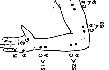
Ulnar to Abd Dig Minimi
|
C8,T1 |
on the ulnar surface of the hypothenar eminence, halfway between the level of the pisiform bone and the 5th metacarpophalangeal joint |
Slightly distal to the 5th metacarpophalangeal joint. |
between the stimulating and recording electrodes |
A- 8 cm proximal to the active electrode, in a line measured slightly radial to the tendon of the flexor carpi ulnaris. B- 4 cm distal to the medial epicondyle. C- 10 cm proximal to stimulation point B D- Axilla, 10 cm proximal to stimulation point C |
3.7 |
6 |
50 |
32 |
|
Ulnar to palmer interosseous
|
C8,T1 |
On the palm, slightly radial to the midpoint of the third metacarpal. |
slightly distal to third MCP joint |
dorsum of the hand |
8 cm proximal to the active electrode, slightly radial to the tendon of the flexor carpi ulnaris. |
4 |
3 |
|
|
|
Ulnar
to 1st dorsal interosseous
|
C8,T1 |
On the dorsum of the first web space, in the center of the triangle formed |
Slightly distal to the fifth MCP joint. |
dorsum of the hand |
8 cm proximal to the active electrode, slightly radial to the tendon of the flexor carpi ulnaris. |
4.7 |
5 |
|
|
|
Radial to Ext Dig Communis
|
C7,8 |
by grasping the radius and ulna of the subject’s pronated forearm with the thumb and middle finger at the junction of the upper third and middle third of the forearm. The index finger is placed halfway between these two points to identify the extensor digitorum communis |
ulnar styloid process |
between the stimulating and recording electrodes |
A- Lateral to the tendon of the biceps at the elbow, as it crosses the flexor crease. B- Axilla |
3.5 |
4.5 |
|
|
|
Radial to Ext Indicis
|
C7,8 |
4 cm proximal to the ulnar styloid, over the motor point of the extensor indicis |
ulnar styloid process |
|
A- 8 cm proximal to the active electrode. B- 8 cm proximal to the lateral epicondyle, over the radial groove. |
2.5 |
1.7 |
|
23 |
|
Suprascapular
to Supra & Infraspinatus
|
C5,6 |
Supraspinatus, 2 cm above the midpoint of the spine of the scapula. Infraspinatus, 2 cm inferior to the midpoint of the spine of the scapula |
On the midline thoracic spine at the same level. |
on the acromion |
Erb’s point |
4.3 4.8 |
1.5 |
|
|
|
Thoracodorsal
to Latissimus
|
C6,7,8 |
On the posterior axillary line at the level of the inferior pole of the scapula. |
On the ipsilateral flank. |
On the ipsilateral lateral chest wall. |
Axilla with arm abducted 90° |
2.7 |
1.5 |
|
|
|
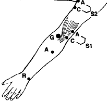
Axillary
|
C5,6 |
most prominent portion of the middle deltoid |
junction of the deltoid muscle and its tendon |
on the acromion |
Erb’s point—the cathode is placed slightly above the upper margin of the clavicle lateral to the clavicular head of the sternocleidomastoid muscle. The anode is superomedial. |
5.4 |
4.6 |
|
|
|
Long
Thoracic
|
C5,6,7 |
A concentric needle electrode (R) is placed at the digitation of the serratus anterior along the midaxillary line over the 5th rib |
|
Anterior axillary line over the 12th rib level |
Erb’s point |
4.8 |
|
|
|
|
Musculocutaneous
to Biceps
|
C5,6 |
just distal to the midportion of the biceps brachii muscle |
Proximal to the antecubital fossa, at junction of the muscle fibers and the biceps tendon |
on the acromion |
Erb’s point |
5.5 |
4 |
|
|
|
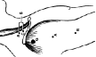
Phrenic
to Diaphragm |
C3,4,5 |
5 cm superior to the tip of the xiphoid process. |
over the upper chest |
|
Stimulation is applied at the posterior border of the sternocleidomastoid muscle in the supraclavicular fossa, with the cathode approximately 3 cm superior to the clavicle. |
8 |
300μ |
|
|