Neuro-Ophthalmology

Quick Facts:

  • The optic nerve head is formed from a coalescence of 1 million axons from the retinal ganglion cells.
  • The optic nerve is divided into the intraocular, intraorbital, intracanicular, and intracranial
  • An altitudinal visual field defect is suggestive of ischemic optic neuropathy but may also be seen in idiopathic optic neuritis.
  • Ischemic optic neuropathy is the result of posterior ciliary artery occlusion.
  • 90% of ischemic optic neuropathy are nonarteritic and related to a congenitally small optic cup and 10% may have underlying giant cell arteritis.
  • more
  • 80% of patients with optic neuritis will have signal intensity abnormality within the optic nerve in STIR sequences with fat-suppressed views
  • In the Optic Neuritis Treatment Trial (ONTT), 59% of patients had cerebral white matter lesions
  • 56% of optic neuritis convert to MS in 10 years if MRI is positive and only 22% if MRI is negative
  • The fibers of the optic tract synapse in: the primary visual pathway synapses in the LGB, the pupillomotor pathway synapses in the pretectum, and the subcortical visual pathway synapses in the superior colliculus
  • Oculomotor complex is located in the dorsal midbrain, anterior to the cerebral aqueduct.
    • The central caudal nucleus innervates both levator muscles
    • The superior rectus receives information from the contralateral superior rectus subnuclei, but the rest of the innervation of the third nerve is ipsilateral.
  • As the oculomotor nerve enters the orbit, it separates into the superior and inferior divisions. The superior division innervates the levator palpebrae and superior rectus, whereas the inferior division innervates the papillary sphincter, medial and inferior recti, and the inferior oblique.
  • A lesion of the third nerve nucleus is exceedingly rare but will result in an ipsilateral third nerve palsy with contralateral superior rectus weakness and bilateral partial ptosis
  • Trochlear nerve floats freely in the cavernous sinus lateral to the internal carotid artery, as opposed to the third and fourth nerves, which are in the lateral wall of the sinus.

.

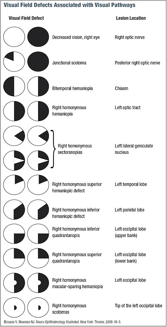
Localization of Visual Field and Higher Cortical Visual Dysfunction

Field Defect or Syndrome Localization
  • Unilateral central scotoma
  • Optic nerve
  • Bitemporal hemianopsia
  • Chiasm
  • Junctional defect (ipsilateral central scotoma and a contralateral superior temporal field cut)
  • Anterior chiasm or posterior optic nerve (Wilbrand knee is affected)
  • Central temporal scotomas
  • Posterior chiasm
  • Incongruous homonymous hemianopsia, afferent pupillary defect, and bow-tie atrophy
  • Optic tract
  • Homonymous sectoranopia
  • Lateral geniculate nucleus
  • Incongruous homonymous hemianopsia
  • Lateral geniculate nucleus
  • Homonymous upper quadrant defect “pie in the sky”
  • Temporal lobe
  • Homonymous defect, denser inferiorly
  • Parietal lobe
  • Gerstmann syndrome and a homonymous defect, denser inferiorly
  • Parietal lobe
  • Complete homonymous hemianopsia
  • Not well-localized to post-chiasmal location
  • Homonymous upper quadrantanopsia with macular sparing
  • Occipital lobe (lower bank)
  • Homonymous lower quadrantanopsia with macular sparing
  • Occipital lobe (upper bank)
  • Isolated homonymous defect (macular sparing) without other neurologic findings
  • Occipital lobe
  • Anton syndrome (cortical blindness)
  • Bilateral occipital lobe lesions
  • Balint syndrome
  • Bilateral occipitoparietal lesions
  • Alexia without agraphia
  • Left occipital lobe and angular gyrus
  • Central achromatopsia
  • Bilateral occipito-temporal lesions


neuro-ophthalmology : visual defects associated with visual pathways

.

Occipital Lobe Lesions:

Syndrome   Localization
  • Anton syndrome
  • Patients with cortical blindness who are unaware of their visual loss
  • Bilateral occipital lesions
  • Palinopsia
  • Perseveration of the visual image once the stimulus has been removed
  • Occipital lobe lesion
  • Prosopagnosia
  • Inability to recognize familiar faces
  • Bilateral occipitotemporal lesions
  • Achromatopsia
  • Abnormality of color perception
  • Bilateral occipitotemporal lesions
  • Balint syndrome
 

  • Triad of simultanagnosia (impaired spatial awareness of more than one object at time), optic apraxia (difficulty in fixating the eyes), and ocular ataxia (visual misreaching)
  • Bilateral parieto-occipital lobes or the visual association cortex
  • Alexia without agraphia
  • left occipital lobe and splenium of the corpus callosum (visual information from the intact right occipital lobe is unable to reach the language areas “angular”)

.

Horner Syndrome Localization:

Associated Symptoms Consideration
  • Isolated, painful
  • Carotid dissection, cluster headache
  • Sensory level
  • Spinal cord
  • Arm numbness or weakness
  • Brachial plexus
  • Ipsilateral face and contralateral body numbness
  • Medulla
  • Sixth-nerve palsy
  • Cavernous sinus

.

Localization of Motility Abnormalities:

Motility Disturbance Localization/Etiology
  • Weber syndrome (Third-nerve palsy and hemiparesis)
  • Anterior midbrain
  • Benedikt syndrome (third-nerve palsy and contralateral tremor)
  • Red nucleus and third-nerve fascicle
  • Isolated pupil-involving third-nerve palsy
  • Posterior communicating artery aneurysm
  • Pupil-sparing third-nerve palsy
  • Microvascular ischemia of the third nerve
  • Isolated fourth-nerve palsy
  • Doral midbrain/anterior medullary velum, Microvascular ischemia
  • Isolated sixth nerve palsy
  • Pons or sixth-nerve fascicle, Demyelination/microvascular ischemia
  • Gaze palsy and facial weakness
  • Dorsal pons/facial colliculus
  • Third-, fourth-, and sixth-nerve palsies
  • Cavernous sinus
  • Third-, fourth-, sixth-nerve palsies, and optic neuropathy
  • Orbital apex
  • Internuclear ophthalmoplegia
  • Medial longitudinal fasciculus
  • Gaze palsy
  • Dorsal pons
  • Parinaud syndrome (upgaze palsy, eyelid retraction)
  • Dorsal midbrain

.

Localization of Pupil Abnormalities:

Abnormality Description Causes
  • Marcus-Gunn pupil (afferent pupillary defect)
  • The affected pupil does not react to light as briskly as the unaffected pupil
  • unilateral optic neuropathies chiasmal and optic tract lesions
  • Horner syndrome
  • Miosis, with an increase in anisocoria in the dark - Ptosis of the upper and lower lids Variable anhydrosis - slow pupillary redilation in the dark
  • Acquired (see above) or Congenital (there will be a heterochromic iris because of impairment of the pigmentary changes.)
  • Adies-Tonic pupil
  • dilated pupil with poor or absent response to light but preserved near response
  • Holmes Adies syndrome.
  • Triad of dilated pupil with poor or absent response to light but preserved near response, loss of deep tendon reflexes, and abnormalities of sweating.
  • Unknown, may be viral infection of parasympathetic and dorsal root ganglia
  • Argyll-Robertson pupils
  • Pupils are small and irregular with impaired light response and intact near response
  • diabetes or syphilis